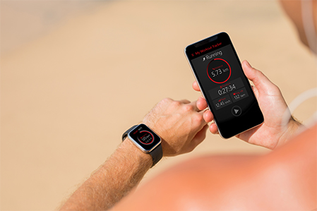
rPPG
Remote photoplethysmography (rPPG) is a non-contact method for measuring physiological signals by analyzing subtle color changes in the skin captured through standard video cameras.1 Like traditional photoplethysmography (PPG), which detects blood volume changes in the microvascular bed of tissue, rPPG does so remotely without physical sensors, registering color changes that occur with each heartbeat. Sophisticated algorithms can then compute vital signs such as heart rate (HR), respiratory rate, and blood oxygen saturation – often by capturing a short video of the face.1
rPPG has attracted growing interest in healthcare2 due to its potential for:
One promising metric measurable via rPPG is heart rate variability (HRV) – the variation in time intervals between heartbeats.3 HRV is a powerful indicator of:
- Autonomic nervous system function – HRV reflects the balance between the sympathetic and parasympathetic nervous systems.
- Stress and fatigue levels – Used in biofeedback techniques to improve stress resilience and emotional regulation. Athletes also use HRV to optimize training and prevent overexertion.
- Cardiovascular health – Low HRV is linked to higher cardiovascular risk and poorer outcomes for patients with existing cardiovascular diseases.
- Early signs of illness or physiological imbalance – Because HRV reflects the body’s ability to adapt to stress and environmental demands, it serves as an indicator of overall health.
rPPG provides a non-invasive, continuous method for measuring HRV and other biomarkers, making it a compelling area of research. Combined with biomarker analysis, it enables health assessments without physical contact or wearable devices.
Accurate measurement using rPPG requires controlled conditions, including consistent lighting, stable camera frame rates, and minimal subject movement. Despite these challenges, advances in computer vision and deep learning are steadily improving the reliability of rPPG-based analysis.
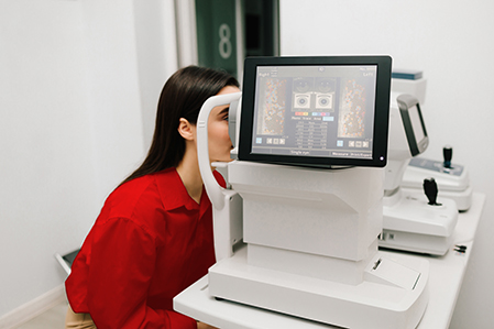
Oculomics
The retinal vasculature mirrors the body’s general circulatory system, while retinal nerve fibers extend directly from the central nervous system. As such, examining the eye, fundus, and retina have long been part of systemic health evaluation.4,5
Advances in digital optics and imaging – including fundus photography, retinal CT, and MR scanning – have enabled wide adoption of non-invasive retinal imaging. Hand-held fundus cameras offer added portability, and high-resolution imaging now reveals retinal biomarkers that were previously invisible to the human eye.
Oculomics – the use of ophthalmic biomarkers to detect, predict, and understand disease – has been made possible by these optical and digital innovations.4,5 Large volumes of retinal images have become fertile ground for AI. Machine learning and deep learning techniques have produced algorithms that accurately detect conditions like diabetic retinopathy, glaucoma, and macular degeneration.6 Some mature models can now make diagnoses without a human specialist, using only high-quality optical images. Achieving sensitivity and specificity of 93% and 91% respectively for diabetic retinopathy detection, FDA approval has been granted to specific AI models.6
Beyond eye disease, researchers are developing retinal biomarkers to detect and predict systemic conditions, including cardiovascular, neurodegenerative, chronic kidney, and hepatobiliary diseases. Researchers are also examining whether specific retinal features correlate with metrics like BMI, blood pressure, cholesterol, and hemoglobin levels – potentially offering a noninvasive alternative to traditional blood tests.4,5
Despite strong momentum, AI models still require validation in large, diverse, real-world populations. Much of the current research remains siloed. Best practice guidelines could help standardize protocols and improve consistency across imaging techniques. Still, even with user acceptance, cost-effectiveness must be demonstrated.
As clinical adoption progresses, the overlap between clinical and insurance objectives may open a nascent path of multi-system risk segmentation through a single non-invasive assessment.