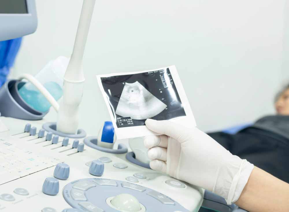
For more than half a century, ultrasound and Doppler have been the two main technologies used to administer screening and diagnostic tests during pregnancy.
These tests provide the fundamental markers and metrics that assess and track fetal health, development, and growth. As ultrasound and Doppler are noninvasive, these can be administered as early as the sixth week of gestation, and are generally done at various time points throughout the nine months. Over the long term, regular ultrasound exams during pregnancy, coupled with health care for both mother and fetus, can contribute to the management of care for mothers and infants by forestalling and limiting prenatal and postpartum complications for both. Strong case management also yields effective cost-of-care management and claims experience, from gestation to birth and beyond.
Background
Fetal medicine encompasses several medical specialties, including cardiology, neurology, maternal-fetal medicine, genetics, and more. Its focus is on optimizing prenatal and postpartum care for both the fetus and mother.
Ultrasound, as a tool for both screening and diagnosis, has become an integral component of fetal medicine. First introduced in the mid-1950s in Scotland, its use has since spread throughout the world. Advances over the years include refinements enabling placenta location and detailed fetal biometry in the 1960s, and the ability to detect fetal weight and intrauterine growth restricted (IUGR) fetuses in the 1970s. Routine ultrasound screening was first introduced in the late 1970s, increasing the identification of pregnancies with fetuses at risk. Transvaginal transducers were introduced in the mid-1980s, providing far better imagery, and Doppler imaging become incorporated around the same time.
Today, doctors start looking for fetal pathologies as early as possible in pregnancy – even as early as Week 6 – and continue to test each trimester in order to ensure development is on target. Ultrasound enables clinicians to study fetal anatomy and physiology in unusually fine detail, detect the manifestations of genetic and developmental abnormalities, and even take corrective steps while the fetus is still developing as well as postpartum.
Ultrasound tests and fetal health
Maternal health is closely linked to fetal and newborn health. According to the World Health Organization (WHO), pregnancy or childbirth complications are the cause of death for more than half a million women worldwide every year (about 830 a day) – complications frequently identifiable via ultrasound. More than 99% of these deaths occur in underdeveloped or developing countries.
WHO also estimates that approximately 15 million babies worldwide (5% to 18% of births) are born prematurely (defined as less than 37 weeks gestation) every year. Preterm birth is the leading cause of prenatal mortality worldwide: the global infant mortality rate, according to the United Nations World Population Progress Report, is 49.4 per 1,000. Again, most are in underdeveloped countries.
Detection of fetal and maternal health conditions early in a pregnancy is vital because it permits the implementation of prevention and surveillance measures, which can improve prognoses, while the lack of such information can result in high economic and quality of life costs. In the U.S., for example, medical costs of caring for preterm babies and babies born with genetic malformations are among the highest-cost claims seen under health policies: RGA has seen claims in the U.S. market for close to US$2 million. While costs in other markets may not be as high, this type of claim can rank among the most expensive.
Timely fetal diagnoses improve fetal and maternal survival rates, strengthen short- and long-term prognoses of fetuses with congenital diseases, manifested genetic abnormalities, and more, and improves prognoses for mothers experiencing health complications.
About the Tests
Testing is important: parents have a right to know the existence of pathology. Early diagnoses can dictate the need for a multidisciplinary management team.
Six different types of ultrasounds are available during a pregnancy:
- Standard. A transducer is placed over the abdomen to generate two-dimensional images of the developing fetus.
- Advanced. Similar to the Standard, the exam uses more sophisticated equipment to target suspected problems.
- Doppler. This technology measures changes in the frequency of waves off moving objects, such as blood cells, and can determine blood flow abnormalities.
- 3-D. This newer technology generates three-dimensional images of a developing fetus.
- 4-D (or dynamic 3-D). This employs scanners that enable a real-time view of a fetus’s face and body movements.
- Fetal Echocardiography. Ultrasound is used to assess a fetus’s heart anatomy and function.
Three to four prenatal ultrasounds can be performed to examine the mother and baby and assess whether development is progressing well. The first can be done at Week 6 or 7 of gestation, the second from Week 11 to 13.6, the third from Week 18 to 23, and the last one from Week 32 to 36.
These ultrasounds produce markers indicative of fetal development, viability, and maturity, as well as fetal and maternal well-being and discomfort. They also examine for fetal pathologies such as genetic abnormalities, cardiac or hemodynamic problems, and for maternal pelvic and uterine abnormalities. For an effective assessment of maternal and fetal health, clinicians should be viewing ultrasound findings integratively, and not in isolation.
- First test (6-7 weeks) confirms pregnancy and determines the due date.
- First trimester tests (11-13.6 weeks) should assess: Fetal biometry, overall fetal size, organ size, presence/absence of nasal bone, fetal heart rate and hemodynamics, and nuchal translucency (thickness of fetal neck).
- Second trimester (weeks 18-23): Detailed examination and tests, including cervical length, growth assessment, sex determination, amniotic fluid volume (testing for oligohydramnios and polyhydramnios), and genetic and other abnormalities and birth defects. It is also when multiples can be determined.
- Third trimester (weeks 32-36) tests: Doppler test for fetal hemodynamics should include umbilical and middle cerebral artery blood flow and fetal cardiac function. Ultrasound test to identify placenta location, observe fetal presentation and movements, identify uterine and pelvic abnormalities, and check maternal uterine arteries for preeclampsia. In addition, mothers should be tested for gestational diabetes.
In certain instances, such as mothers being over age 35 and/ or having gestational diabetes, or cases of placenta previa (where the placenta is covering the cervix), testing can be more frequent. Doctors will sometimes order additional ultrasounds closer to the delivery date to ensure the health of the mother and fetus. Early recognition of a risky condition enables medical specialist teams to be organized to reduce maternal and fetal mortality and future neonatal disabilities, and get better control over the cost of care.
What insurers can do
Effective pregnancy management reduces the healthcare burden for both the insured and the insurer. It further helps to provide a better quality of life for the infant, the mother, and the family. Insurance companies could play a more active role in supporting the policyholders during the gestation period: providing information and advice to pregnant mothers, and developing programs to identify and monitor mothers at high risk of premature delivery. In addition, product designs should ensure the level of maternity cover is sufficient to allow for monitoring and treatment of any complication.
From a claims point of view, processes, and controls should be in place to prevent abuse, along with having strong provider agreements to manage costs.
Conclusions
Ultrasonography has produced spectacular improvements in fetal diagnostics and care. The markers it can generate in each stage of pregnancy are of great importance, as timely information about maternal and fetal health can provide valuable knowledge and contribute to successful pregnancies. Insurance companies could play a bigger role in informing and encouraging insureds about how ultrasounds can screen for, diagnose, and possibly prevent complications in pregnancy.